Pages

Saturday, May 7, 2016

HydroFlex™ microplate washer for 96-well format


- Download Brochure 
 حمل كتالوج الجهاز من اللينك الاتي

The HydroFlex microplate washer is a truly flexible platform that provides excellent automated microplate strip washing and vacuum filtration performance for 96-well microplate formats.
This modular and upgradeable platform is ideal for a wide range of cell-, enzyme- and DNA-based applications in academia, biotech, pharma and clinical diagnostics, reflecting over 30 years of Tecan expertise in advanced liquid handling.

Integrated application flexibility

The multifunctional HydroFlex microplate washer offers far greater flexibility than a standard plate microplate washer, and features user-interchangeable plate carriers for easy switching between washing and filtration applications to allow:
  • ELISA washing for improved consistency and reliability
  • Gentle washing of cell-based assays with adherent or weakly adherent cells
  • Microplate array washing for protein and antibody arrays
  • Vacuum filtration to waste for PCR cleanup after DNA amplification, etc.
  • Magnetic bead washing using MBS carrier
HydroFlex features and benefits:
  • Compact and modular design, suitable for a range of applications and laboratories
  • Ready for automated washing of ELISAs, cells and protein arrays
  • Automated system for vacuum filtration to waste
  • Magnetic plate carrier for magnetic bead washing
  • Advanced online process controls for safety and reliability
  • Bubble sensor unit that ensures reliable buffer dispensing
  • Multipoint aspiration for flat bottom plates to achieve minimal residual volumes
  • Individual software control of speed settings and wash head positions
  • Suitable for integration onto Freedom EVO and Freedom EVOlyzer workstations
  • Excellent reliability and low service costs

Thursday, May 5, 2016

Different Size, Shape and Arrangement of Bacterial Cells

Different Size, Shape and Arrangement of Bacterial Cells


- برجاء قراءة الصفحة الى اخرها لمعرفة اجهزة الاليزا وPCR و أجهزة تحليل CBC واجهزة تحليل الهرمونات ودلالات الاورام
Bacteria are prokaryotic, unicellular microorganisms, which lack chlorophyll pigments. The cell structure is simpler than that of other organisms as there is no nucleus or membrane bound organelles.
Due to the presence of a rigid cell wall, bacteria maintain a definite shape, though they vary as shape, size and structure.


When viewed under light microscope, most bacteria appear in variations of three major shapes: the rod (bacillus), the sphere (coccus) and the spiral type (vibrio). In fact, structure of bacteria has two aspects, arrangement and shape. So far as the arrangement is concerned, it may Paired (diplo), Grape-like clusters (staphylo) or Chains (strepto). In shape they may principally be Rods (bacilli), Spheres (cocci), and Spirals (spirillum).

Size of Bacterial Cell
 
Size of Bacterial Cells

The average diameter of spherical bacteria is 0.5-2.0 µm. For rod-shaped or filamentous bacteria, length is 1-10 µm and diameter is 0.25-1 .0 µm.
  • E. coli , a bacillus of about average size is 1.1 to 1.5 µm wide by 2.0 to 6.0 µm long.
  • Spirochaetes occasionally reach 500 µm in length and the cyanobacterium
  • Oscillatoria is about 7 µm in diameter.
  • The bacterium, Epulosiscium fishelsoni , can be seen with the naked eye (600 mm long by 80 mm in diameter).
  • One group of bacteria, called the Mycoplasmas, have individuals with size much smaller than these dimensions. They measure about 0.25 µ and are the smallest cells known so far. They were formerly known as pleuropneumonia-like organisms (PPLO).
  • Mycoplasma gallicepticum, with a size of approximately 200 to 300 nm are thought to be the world smallest bacteria.
  • Thiomargarita namibiensis is world’s largest bacteria, a gram-negative Proteobacterium found in the ocean sediments off the coast of Namibia. Usually it is 0.1—0.3 mm (100—300 µm) across, but bigger cells have been observed up to 0.75 mm (750 µm).
Thus a few bacteria are much larger than the average eukaryotic cell (typical plant and animal cells are around 10 to 50 µm in diameter).
Shape of Bacterial Cell 

The three basic bacterial shapes are coccus (spherical), bacillus (rod-shaped), and spiral (twisted), however pleomorphic bacteria can assume several shapes.

Shape of Bacterial Cell


  • Cocci (or coccus for a single cell) are round cells, sometimes slightly flattened when they are adjacent to one another.
  • Bacilli (or bacillus for a single cell) are rod-shaped bacteria.
  • Spirilla (or spirillum for a single cell) are curved bacteria which can range from a gently curved shape to a corkscrew-like spiral.  Many spirilla are rigid and capable of movement.  A special group of spirilla known as spirochetes are long, slender, and flexible.


Arrangement of Cocci
 
Cocci bacteria can exist singly, in pairs (as diplococci ), in groups of four (as tetrads ), in chains (as streptococci ), in clusters (as stapylococci ), or in cubes consisting of eight cells (as sarcinae). Cocci may be oval, elongated, or flattened on one side. Cocci may remain attached after cell division. These group characteristics are often used to help identify certain cocci.

1. Diplococci

The cocci are arranged in pairs.
Examples: Streptococcus pneumoniae, Moraxella catarrhalis, Neisseria gonorrhoeae, etc.

Diplococci and Streptococci


2. Streptococci

The cocci are arranged in chains, as the cells divide in one plane.
Examples: Streptococcus pyogenes, Streptococcus agalactiae


3. Tetrads

Tetrads
The cocci are arranged in packets of four cells, as the cells divide in two plains.
Examples: Aerococcus, Pediococcus and Tetragenococcus

4. Sarcinae

Sarcinae

The cocci are arranged in a cuboidal manner, as the cells are formed by regular cell divisions in three planes. Cocci that divide in three planes and remain in groups cube like groups of eight.
Examples: Sarcina ventriculi, Sarcina ureae, etc.

5. Staphylococci

Staphylococci

The cocci are arranged in grape-like clusters formed by irregular cell divisions in three plains.
Examples: Staphylococcus aureus

Arrangement of Bacilli
 
The cylindrical or rod-shaped bacteria are called ‘bacillus’ (plural: bacilli).

1. Diplobacilli

Diplobacilli

Most bacilli appear as single rods. Diplobacilli appear in pairs after division.
Example of Single Rod: Bacillus cereus Examples of Diplobacilli: Coxiella burnetii, Moraxella bovis, Klebsiella rhinoscleromatis, etc.

2. Streptobacilli

Streptobacilli

The bacilli are arranged in chains, as the cells divide in one plane.
Examples: Streptobacillus moniliformis

3. Coccobacilli

Coccobacilli

These are so short and stumpy that they appear ovoid. They look like coccus and bacillus.
Examples: Haemophilus influenzaeGardnerella vaginalis, and Chlamydia trachomatis

4. Palisades

Palisades

The bacilli bend at the points of division following the cell divisions, resulting in a palisade arrangement resembling a picket fence and angular patterns that look like Chinese letters.
Example: Corynebacterium diphtheriae 

Arrangement of Spiral Bacteria
 
Spirilla (or spirillum for a single cell) are curved bacteria which can range from a gently curved shape to a corkscrew-like spiral.  Many spirilla are rigid and capable of movement.  A special group of spirilla known as spirochetes are long, slender, and flexible.

1. Vibrio

Vibrio

They are comma-shaped bacteria with less than one complete turn or twist in the cell.
Example: Vibrio cholerae

2. Spirilla

Spirilla

They have rigid spiral structure. Spirillum with many turns can superficially resemble spirochetes. They do not have outer sheath and endoflagella, but have typical bacterial flagella.
Example: Campylobacter jejuni, Helicobacter pyloriSpirillum winogradskyi, etc.

3. Spirochetes

Spirochetes

Spirochetes have a helical shape and flexible bodies. Spirochetes move by means of axial filaments, which look like flagella contained beneath a flexible external sheath but lack typical bacterial flagella.
Examples: Leptospira species (Leptospira interrogans), Treponema pallidumBorrelia recurrentis, etc.

Others Shapes and Arrangements of Bacteria

1. Filamentous Bacteria

Filamentous bacteria

They are very long thin filament-shaped bacteria. Some of them form branching filaments resulting in a network of filaments called ‘mycelium’.
Example: Candidatus Savagella

2. Star Shaped Bacteria

Star Shaped Bacteria
Example: Stella

3. Rectangular Bacteria

Rectangular Bacteria

Examples: Haloarcula spp (H. vallismortis, H. marismortui)

4. Pleomorphic Bacteria

Pleomorphic Bacteria

These bacteria do not have any characteristic shape unlike all others described above. They can change their shape. In pure cultures, they can be observed to have different shapes.
Examples: Mycoplasma pneumoniae, M. genitalium, etc.

لمعرفة المواصفات وتحميل الكتالوج اضغط علي صورة كل جهاز


لمعرفة المواصفات وتحميل الكتالوج اضغط علي صورة كل جهاز

 جهاز الاليزا  

لمعرفة المواصفات وتحميل الكتالوج اضغط علي صورة كل جهاز

  أفضل الاسعار لجهاز عد خلايا الدم السويدى الصنع

لمعرفة المواصفات وتحميل الكتالوج اضغط علي صورة كل جهاز

  جهاز الهرمونات ودلالات الاورام الالمانى الصنع


Wednesday, May 4, 2016

Types of Crystals in Urine

 جهاز الاليزا  جهاز  Real-Time PCR
 CBC جهاز تحليل   جهاز الهرمونات ودلالات الاورام


Types of Crystals in Urine

Uric acid crystals invariably form in acidic urine, typically with a urine pH < 5.5. Uric acid is soluble in alkaline urine, preventing the precipitation of urate crystals. The inability of uric acid to crystallize at urine pH > 7.0 is the rationale for urinary alkalinization in patients at risk for acute uric acid nephropathy. Uric acid crystalluria is not associated with significant amounts of hematuria, glycosuria or proteinuria.


Although crystals can be seen in certain clinical scenarios, such as kidney stone disease or acute crystal nephropathy, visualizing crystals under the microscope does not guarantee that the crystals were present in the urinary system. Crystals can continue to form after micturition. Crystal precipitation after micturition is most commonly due to changes in temperature, as can occur if the urine is stored at room temperature or in a refrigerator, or changes in urinary pH, as can occur in the presence of infection due to urea-splitting organisms.
 ـــــــــــــــــــــــــــــــــــــــــــــــــــــــــــــــــــــــــــــــــــــــــــــــــــــــــــــــــــــــــــــــ

Urate crystals

Urate Crystals 

Uric acid crystals can vary in both size and shape, as can be seen in the slide above. They can look like barrels, rosettes, rhomboids, needles or hexagonal plates. They are usually amber in color, irrespective of the size or shape of the individual crystal. However, urate crystals may assume the color of any pigments (such as bilirubin or the medication pyridium) that are present in the urine. Urate crystals can occasionally be seen in normal subjects, although they are much more common in patients with urate nephrolithiasis or acute urate nephropathy.

 ـــــــــــــــــــــــــــــــــــــــــــــــــــــــــــــــــــــــــــــــــــــــــــــــــــــــــــــــــــــــــــــــ

Cystine Crystals

Cystine Crystals

In contrast to polymorphic urate crystals, cystine crystals are monomorphic, colorless hexagonal plates which look similar to benzene rings. The urine sediments from two patients with cystine crystals are shown above. Cystine crystals may be isolated or may be heaped upon one another. They occur in the sediment of patients with cystinuria, a genetic defect in renal cystine transport. They are found in acidic urine, typically with a urine pH < 6.0.
 ـــــــــــــــــــــــــــــــــــــــــــــــــــــــــــــــــــــــــــــــــــــــــــــــــــــــــــــــــــــــــــــــ

Triple phosphate, or struvite
 
Triple phosphate, or struvite

Triple phosphate, or struvite, crystals are described as having a “coffin-lid”-shaped appearance. Several struvite crystals are shown above. Struvite crystals are composed of magnesium ammonium phosphate. They are typically seen in alkaline urine, with a urine pH > 7.0. Triple phosphate crystals are seen in patients with urinary tract infections caused by urea-splitting bacteria, such as Proteus mirabilis, and are frequently found in the urine of patients with infected calculi (struvite stones). In addition to triple phosphate crystals, microscopy in these patients with urinary tract infections may show significant leukocyturia (arrows, above right) and bacteriuria.
 ـــــــــــــــــــــــــــــــــــــــــــــــــــــــــــــــــــــــــــــــــــــــــــــــــــــــــــــــــــــــــــــــ

Calcium oxalate crystals
 
Calcium oxalate crystals

Calcium oxalate crystals are usually found in acidic urine. They may occur as either bihydrated or monohydrated calcium oxalate. Calcium oxalate bihydrate crystals appear as colorless bipyramids of various sizes (“envelope form”, above left). Calcium oxalate monohydrate crystals are colorless and can assume several shapes, including ovoids, biconcave disks, rods and dumbbells (above right, yellow arrows). They can be seen in normal individuals with high dietary oxalate ingestion, in patients with nephrolithiasis, and in patients with acute renal failure due to ethylene glycol ingestion.
 ـــــــــــــــــــــــــــــــــــــــــــــــــــــــــــــــــــــــــــــــــــــــــــــــــــــــــــــــــــــــــــــــ

Calcium carbonate crystals 
 
Calcium carbonate crystals

Calcium carbonate crystals are variably sized crystals that frequently appear as large spheroids with radial striations. They can also be seen as smaller crystals with round to ovoid shapes. they are colorless to yellow-brown and can impart a brownish tinge to the urine, when they occur in high numbers. They are usually large crystals and can be readily observed at low magnification (however, confirmation of crystal identity should always be performed under high magnification and smaller variants of calcium carbonate may be missed if only low magnification is used). These crystals are common in the urine of normal horses, rabbits, guinea pigs and goats. They have not been observed in canine or feline urine.
 ـــــــــــــــــــــــــــــــــــــــــــــــــــــــــــــــــــــــــــــــــــــــــــــــــــــــــــــــــــــــــــــــ

“Amorphous” crystals
 
Amorphous crystals

“Amorphous” crystals appear as aggregates of finely granular material without any defining shape at the light microscopic level. They can be comprised of urates, phosphates or xanthine. They are usually small crystals that are usually only observed at high magnification (unless there are large amounts of them), i.e. they mimic bacteria.
Amorphous urates (Na, K, Mg, or Ca salts) tend to form in acidic urine and may have a yellow or yellow-brown color. Amorphous phosphates are similar in general appearance, but tend to form in alkaline urine and lack color. Xanthine crystals are usually in the form of “amorphous” crystals. These crystals occur in Dalmations on allopurinol therapy for urate urolithiasis. Generally, no specific clinical interpretation can be made based on the finding of amorphous crystals. Small amorphous crystals can be confused with bacterial cocci in some cases, but can be distinguished by gram-staining. Degenerating crystals or cells can also resemble “amorphous” crystals.
 ـــــــــــــــــــــــــــــــــــــــــــــــــــــــــــــــــــــــــــــــــــــــــــــــــــــــــــــــــــــــــــــــ

Ammonium biurate
 
Ammonium biurate crystals

Ammonium urate (or biurate) crystals generally appear as brown or yellow-brown spherical bodies with irregular protrusions (“thorn-apples”). In some urine samples, they do not have irregular protrusions but have smooth borders and can resemble calcium carbonate (although these do not occur in the urine from dogs and cats). These crystals can be observed under low magnification, particularly when seen in large number, however low numbers may only be seen at higher magnification. Due to their potential pathologic relevance, crystal identification should be verified by examination at high magnification. Though possible in urine of any pH, their formation is favored in neutral to alkaline urine. They are frequently seen with amorphous urates. These crystals are fairly common in dogs and cats with congenital or acquired portal vascular anomalies, with or without concomitant ammonium urate uroliths. They can be seen in urine from normal Dalmatians and Bulldogs, both of which .are predisposed to urate urolithiasis. They are rarely, if ever, seen in urine from normal cats or dogs of other breeds and have not been reported in large animals.
 ـــــــــــــــــــــــــــــــــــــــــــــــــــــــــــــــــــــــــــــــــــــــــــــــــــــــــــــــــــــــــــــــ

Bilirubin crystals
 
Bilirubin crystals

Bilirubin crystals form from conjugated bilirubin (water soluble) and are needle-like to granular crystals that are yellow in color. They tend to precipitate onto other formed elements in the urine. In the top picture, fine needle-like crystals have formed on an underlying cell. This is the most common appearance of bilirubin crystals. In the lower two pictures, cylindrical bilirubin crystals have formed in association with droplets of fat, resulting in a “flashlight” appearance. This form is less commonly seen. They are usually small crystals that are usually only observed at high magnification (unless there are large aggregates of crystals). Bilirubin crystals are seen most commonly in canine urine, especially in highly concentrated specimens. They are less common in urine of other species. In dogs, they often are of no clinical significance (healthy dogs can have low, but detectable, bilirubin levels in urine). Bilirubin crystals (or a positive chemical reaction on the urine dipstick) in feline, equine, bovine, or camelid urine is an abnormal finding and the animal should be investigated for an underlying cholestatic process.
 ـــــــــــــــــــــــــــــــــــــــــــــــــــــــــــــــــــــــــــــــــــــــــــــــــــــــــــــــــــــــــــــــ

Sulfadiazine crystal, Indinavir crystal
 
sulfadiazine crystal 

indinavir crystal
Urinary crystals can also be seen in patients taking certain medications. One example is sulfadiazine: these crystals appear as striated shells or “shocks of wheat.“ A sulfadiazine crystal is shown on the left. Other medications that can cause urine crystals to form include indinavir, intravenous acyclovir, and triamterene. When these medications are given in high doses or to volume depleted patients, the crystals can cause acute renal failure by crystalline blockage of the renal tubules. An example of an indinavir crystal is shown on the right: the top panel shows rectangular plates of various sizes containing needle crystals, while the bottom panel shows indinavir crystals in a sheaf of numerous, densely packed needles.
 ـــــــــــــــــــــــــــــــــــــــــــــــــــــــــــــــــــــــــــــــــــــــــــــــــــــــــــــــــــــــــــــــ
 جهاز الاليزا  جهاز  Real-Time PCR

 CBC جهاز تحليل   جهاز الهرمونات ودلالات الاورام


Differences between Gram Positive and Gram Negative Bacteria


Differences between Gram Positive and Gram Negative Bacteria


S.N.

Characteristics

Gram Positive

Gram Negative

1
Gram Reaction
Gram Positive StainRetain crystal violet dye and stain blue or purple
Gram Negative StainCan be decolorized to accept counterstain (safranin) and stain pink or red
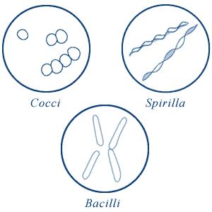
2
Cell Wall
Cell Wall is 20-30 nm thick.
Cell Wall is 8-12 nm thick.
3
Cell Wall
The wall is Smooth.
The wall is wavy.
4
Peptidoglycan Layer
Thick (multilayered)
Thin (single-layered)
5
Teichoic Acids
Present in many
Absent
6
Periplasmic Space
Absent
Present
7
Outer Membrane
Absent
Present
8
Porins
Absent
Occurs in Outer Membrane
9
Lipopolysaccharide (LPS) Content
Virtually None
High
10
Lipid and Lipoprotein Content
Low (acid-fast bacteria have lipids linked to peptidoglycan)
High (because of presence of outer membrane)
11
Mesosomes
Quite Prominent
Less Prominent
12
Flagellar Structure
2 rings in basal body
4 rings in basal body
13
Toxin Produced
Exotoxins
Endotoxins or Exotoxins
14
Resistance to Physical Disruption
High
Low
15
Cell Wall Disruption by Lysozyme
High
Low (requires pretreatment to destabilize outer membrane)
16
Susceptibility to Penicillin and Sulfonamide
High
Low
17
Susceptibility to Streptomycin, Chloramphenicol and Tetracycline
Low
High
18
Inhibition by Basic Dyes
High
Low
19
Susceptibility to Anionic Detergents
High
Low
20
Resistance to Sodium Azide
High
Low
21
Resistance to Drying
High
Low

Differences between Gram Positive and Gram Negative Bacteria